Early microscopy
The first optical microscope and its inventor are not known, but an early version was produced in 1590 by one of two commonly credited eye glass makers, Hans Lippershey and Zacharias Janssen in the Netherlands. The name "microscope" is attributed to Giovanni Faber in reference to Galileo Galilei's compound microscope in 1625.
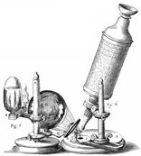
Probably the most well know early microscopy study is that portrayed in Robert Hooke's Micrographia, published in 1665. Hooke was the first person to refer to biological cells and used a compound microscope as shown in the image to the left.
Probably the most well know early microscopy study is that portrayed in Robert Hooke's Micrographia, published in 1665. Hooke was the first person to refer to biological cells and used a compound microscope as shown in the image to the left.
|
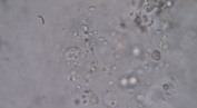
Antonie van Leeuwenhoek is probably one of the most significant biologist of the early microscopists. He founded microbiology by being the first person to observe and report single celled micro-organisms (1676). Among his discoveries are protists, bacteria and spermatozoa.
|
|
The Start of Electron Microscopy
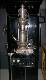
Using a beam of electrons rather than light and electromagnets instead of glass lenses, a leap in the resolution capabilities of microscopes was made in the 1930's. Max Knoll and Ernst Ruska built the first electron microscope in 1931, achieving a resolution greater than possible with light microscopy in 1933. The first electron microscopes were transmission electron microscopes (TEM) and they became commercially available in 1939.
Scanning electron microscopy was demonstrated by Max Knoll in 1935 but was not commercially available until 1965when they were sold by the Cambridge Scientific Instrument Company as the Stereoscan.
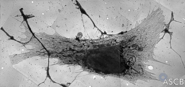
"A Study of Tissue Culture Cells by Electron Microscopy," by Keith Porter, Albert Claude, and Ernest Fullam published in the Journal of Experimental Medicine, 1945, showed the first electron micrograph of an intact cell (shown below as an image montage). This began the science of biological electron microscopy, which has been expanded upon ever since.
Scanning electron microscopy was demonstrated by Max Knoll in 1935 but was not commercially available until 1965when they were sold by the Cambridge Scientific Instrument Company as the Stereoscan.
"A Study of Tissue Culture Cells by Electron Microscopy," by Keith Porter, Albert Claude, and Ernest Fullam published in the Journal of Experimental Medicine, 1945, showed the first electron micrograph of an intact cell (shown below as an image montage). This began the science of biological electron microscopy, which has been expanded upon ever since.